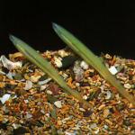
Lancelets(lat. Branchiostoma) - genus primitive marine animals from the lancelet family, subtype skullless (lat.Acrania), class cephalochordates. Adult individuals lead a benthic lifestyle - they inhabit the sandy bottom of clean sea waters; larvae are plankton in coastal areas and the open sea.
A typical representative of the genus is the European lancelet. Considered as an intermediate link between vertebratesand invertebrates animals.
Structure
Appearance
The body of lancelets is translucent, whitish to creamy yellow, sometimes with a shade of pink, with a faint metallic sheen, laterally compressed and elongated. It is pointed at the rear end, and obliquely cut at the front, the ventral side is slightly wider than the dorsal side. The body length of lancelets varies between 5-8 cm.
On the underside of the anterior end of the animal there is a pre-oral funnel (or cavity) surrounded by oral tentacles.
A fin fold stretches along the entire back - a low dorsal fin. It is transparent and supported by numerous rod-shaped fin rays. The dorsal fin, without a visible border, becomes caudal, spear-shaped or lanceolate. The caudal fin functions as propulsion. On the ventral side, along the lower edge of the tail, there is a short subcaudal fin (also erroneously called the ventral fin). The border of the caudal and subcaudal fins is marked by the anus. All these fins, oriented in a plane of bilateral symmetry, act as stabilizers when moving, that is, they prevent the animal from turning over.
From the anterior end of the body to the subcaudal fin, so-called metapleural folds run along the sides. At the point of convergence of the metapleural folds and the subcaudal fin there is an atriopore, or gill pore, the outlet of the atrial, or circumbranchial, cavity.
Skin covering
The skin of lancelets is a single-layer epithelium (epidermis), which is located on the underlying thin basement membrane. On top, the epidermis is covered with a cuticle, a surface film of mucopolysaccharides secreted from the epidermal glands; it protects the thin skin of lancelets from damage. Under the epithelium there is a thin layer of gelatinous connective tissue - corium, or cutis. The outer covers are transparent, almost not pigmented.
central nervous system
The central nervous system (CNS) of lancelets is the neural tube, lying above the chordwith a narrow cavity inside - the neurocoelium. The anterior end of the neural tube is shorter than the chord, this feature gave the name to the subtype - cephalochordates. Head and back are not externally differentiated, but the head and dorsal parts of the neural tube have a distinct structure and perform different functions:
- The head end of the neural tube, approximately two muscle segments long, is responsible for regulating reflex activity. Destruction of this section causes loss of coordination. The nerve coelus widens slightly in this section. This expansion is considered the rudiment, or rudiment, of the cerebral ventricle (brain vesicle). In lancelet larvae, this cavity is connected by an opening (neuropore) to the olfactory organ lying on the surface of the body - Kölliker's fossa, and does not communicate with the environment. Two pairs of sensory head nerves, responsible for the innervation of the head end of the body, depart from the cerebral vesicle. Clusters of special ganglion cells (multipolar neurons) are observed, and in the anterior part of the neural tube there is an unpaired eye, a pigment spot, the function of which has not yet been clarified; it is possible that this is a remnant of the organ of balance.
- Two pairs (right and left) of spinal nerves depart from the dorsal end of the neural tube: dorsal in the anterior part of each segment and ventral in its posterior part. The abdominal motor nerve, with a base of several roots, branches in the myomere. The motor-sensory spinal nerve begins with one root and includes sensory fibers (mainly in the skin) and motor fibers (in muscle tissue). Here, the difference between lancelets (like all skullless animals) from higher chordates is that the dorsal and ventral roots are not united into a single nerve. The position of the nerves is identical to the location of the right and left myomeres, that is, they are also characteristically shifted.
The right and left sides of each segment of the neural tube are connected by nerve cells that form reflex arcs and neurons.
Since rudiments are present in the head section of the neural tube, it can be assumed that the central nervous system of the modern lancelet is more primitive than that of its ancestors, this can be associated with the more active lifestyle of the latter.

Sense organs
The senses are primitive.Tactile sensations are perceived by the nerve endings of the entire epidermis, especially by the oral tentacles. Chemical stimuli are perceived by encapsulated nerve cells, which are also found in the skin and line the Kölliker fossa. In the neural tube, mainly in the area of its cavity, there are light-sensitive cells with concave pigment cells - the ocelli of Hesse. The translucent covers of the animal freely transmit light rays, which are captured by Hessian eyes. They also work like photo relays, recording the position of the lancelet's body in the substrate.
Skeleton
The axial skeleton of lancelets is the notochord, or notochord. This is a light, vertically striated rod that stretches, becoming thinner, along the dorsal side of the body from the anterior end to the posterior. In lancelets, the notochord extends deep forward into the cephalic end, beyond the neural tube.
The notochord of the lancelet, together with the neural tube, is surrounded by a connective tissue membrane, to which the partitions between the myomeres, the myosepta, are connected.
Digestive system
On the lower part of the head end there are oral tentacles and a pre-oral funnel leading into a small oral opening. It is surrounded by a muscular annular membrane - a sail. The sail acts as a partition between the mouth opening and the large pharynx. The front part of the sail is covered with thin ribbon-like outgrowths of the ciliated organ, the back part has short tentacles directed into the pharynx cavity; they are an obstacle to large food particles.
The pharynx in lancelets occupies up to a third of the body length and is penetrated by gill slits with over 100 pairs. The gill slits are separated by interbranchial septa with ciliated epithelium and lead into the peripharyngeal, or atrial cavity, and not directly out. (Gill slits are not visible from the outside; they are covered with protective skin folds.)
The atrial cavity surrounds the pharynx on the sides and below and has an opening that opens outwards - the atriopore. In the form of a blind closed outgrowth, the atrial cavity extends slightly beyond the atriopore. The movements of the outgrowths of the ciliated organ and the vibrations of the cilia covering the interbranchial septa direct a slow and non-stop flow of water into the pharynx. Next, the water passes through the gill slits into the peribranchial cavity, and from there it is removed through the atriopore.
The pharynx has two grooves lined with ciliated and glandular epithelium. The subbranchial groove (subbranchial groove, endostyle) runs along the lower part of the pharynx, and the epibranchial groove (suprabranchial groove) runs along the dorsal side of the pharynx. They are connected by two strips of ciliated epithelium, which run along the lateral inner surfaces of the pharynx in its anterior part. The cells of the endostyle secrete mucus, which, under the influence of the flickering of the cilia, is driven towards the anterior end of the pharynx - towards the flow of water. Along the way, food that enters the pharynx is enveloped and captured. After this, the lumps of food glued together by mucus move along two semicircular grooves into the epibranchial groove, along which they are driven back to the initial section of the intestine (intestines). The mucus in which the food was wrapped flows down the sides of the pharynx and forms a mucous membrane on the gill slits that allows water to pass out.
Sharply narrowing, the pharynx passes into a short, without bends, intestine, which ends in the anus. At the junction of the pharynx and the intestine there is a blind finger-shaped hepatic outgrowth that secretes digestive enzymes. It is located on the right side of the pharynx and is directed towards the head end of the lancelet. Digestion occurs both in the cavity of the hepatic outgrowth and throughout the intestine.
Excretory system
The excretory system of lancelets is compared with the nephridial system of annelids and flat worms It is something between protonephridialand metanephridial system.
About 100 pairs of nephridia metamerically located above the pharyngeal cavity. They look like a short, steeply curved tube that opens into the atrial cavity. Almost the entire rest of the nephridia enters as a whole (supraglottic canals). This part of the tube has nephrostomes - a few openings closed by a group of solenocytes, specialized cells with a “flickering flame” - a constantly working flagellum. Capillary glomeruli are adjacent to the walls of the nephridium tube, through the walls of which metabolic products enter as a whole. From the coelom, decay products penetrate into the solenocyte, and from there into the lumen of the nephridial tube, through which they move with the help of the beating of the flagella of the solenocytes and the ciliated epithelial cells lining the tube. From there, through the opening of the nephridium, waste enters the peribranchial cavity and is removed from the body of the lancelet.
In addition to the nephridia serially located in each metamer, the lancelet has an unpaired (left) Hatchek nephridia, which is the first to appear in ontogenesis. In its structure it resembles other nephridia.
For decades, the origin of lancelet protonephridia remained unclear. Old authors (Goodrich and others) were inclined to believe that they had an ectodermal origin (for example, Goodrich described their development from single-celled rudiments, which, in his opinion, belonged to ectomesoderm). Thus, it was assumed that lancelet nephridia are not homologous to the mesodermal nephrons (kidneys) of vertebrates. Recently, molecular biological data have been accumulating to support the mesodermal origin of lancelet nephridia
Respiratory system
The respiratory system is characterized by the fact that there are no specialized organs. Gas exchangeproduced across the entire surface of the body.

Circulatory system
The circulatory system is closed and delimited from surrounding organs by the walls of blood vessels.
Below the pharynx is the abdominal aorta, a large vessel whose walls constantly pulsate and transport blood, thus replacing the heart. The pulsation occurs through a slow, uncoordinated contraction of the myoepithelial layer of adjacent coelomic cavities. Through the abdominal aorta, venous blood moves to the head end of the body. Through the thin covers of hundreds of gill arteries (efferent), extending according to the number of interbranch septa from the abdominal aorta, oxygen dissolved in water is absorbed by the blood. The bases of the gill arteries - the bulbs - also have the ability to pulsate. The branchial arteries flow into the paired (right and left) roots of the dorsal aorta, which is located at the posterior edge of the pharynx and extends under the notochord to the end of the tail. The anterior end of the body is supplied with blood by two short branches of the paired roots of the dorsal aorta - the carotid arteries. Arteries branching from the dorsal aorta carry blood to all parts of the body. This is how the arterial circulatory system of lancelets is represented.
Having passed through the capillary system, venous blood from the intestinal walls is collected in the azygos intestinal vein, which runs in the form of the hepatic vein to the hepatic outgrowth. In it, the blood again crumbles into capillaries - the portal system of the liver is formed. The capillaries of the hepatic process again merge into a short hepatic vein, which flows into a small extension - the venous sinus. From both ends of the body, blood collects in paired anterior and posterior cardinal veins. On each side they merge and form the right and left ducts of Cuvier (common cardinal veins), which flow into the sinus venosus, which is the beginning of the abdominal aorta. It follows from this that lancelets have one circle of blood circulation. Their blood is colorless and does not contain respiratory pigments. The oxygen saturation of blood in arteries and veins is similar - the small size of animals and single-layer skin make it possible to saturate the blood with oxygen not only through the gill arteries, but through all the superficial vessels of the body.
Reproductive system
Representatives of the genus Lancelets, like other skullless, dioecious: each animal develops either ovaries or testes. The gonads of males and females are similar in appearance - they are spherical protuberances distributed segment by segment on the body wall adjacent to the atrial cavity. Gonadsusually 25-26 pairs. There are no reproductive ducts, and mature germ cellsenter the atrial cavity through ruptures in the walls of the gonads and body walls ; with the flow of water through the atriopore they are released into the external environment. Immature lancelets do not have genital organs.
Lifestyle and nutrition
They live in many seas of the tropical and temperate zones, including the Black Sea, in coastal areas with a clean sandy bottom. Since lancelets are benthic animals, they spend most of their time at the bottom, taking different feeding positions depending on the looseness of the sand. If the soil is loose, lancelets burrow deeply into the substrate and expose only the front end of the body; if the soil is muddy and dense, the animals lie on the bottom. When disturbed, lancelets can swim a short distance and burrow again or lie down on the ground. They can also move through wet sand.
Lancelets live in colonies of more than nine thousand individuals per square meter. They make seasonal migrations - they swim several kilometers.
Lancelets are filter-feeding animals: food is absorbed through the mouth opening with a flow of water, which is driven by the movement of the cilia. The food for lancelets is mainly phyto- and zooplankton - various cladocerans, ciliates, diatoms; as well as larvae and eggs of other lower chordates and invertebrates. The nature of nutrition is passive.
Reproduction
Lancelets reproduce in spring, summer or autumn. Immediately after sunset, females begin to lay mature eggs (eggs). Fertilization occurs in water, as does the subsequent individual development of lancelets.
The embryonic development of lancelets is used in many textbooks and manuals as an example to describe the embryogenesis of chordates, as it represents a simplified diagram of the development of all higher chordates.
Information taken from the sitewww.wikipedia.org
Studying the object
Examine the external structure of the lancelet using a magnifying glass on whole adult fixed specimens.
The lancelet lives in the coastal strip of the seabed. Usually it lies on the ground. The body length is 3-8 cm. It buries itself in the sand with its rear end. Body color is whitish. The ventral side is wider, and the dorsal side is narrow. Toward the rear end, the entire body is pointed in the shape of a lancet, hence the name of the animal. The dorsal fin bends around the rear end of the body and forms the caudal fin and passes on the ventral side into a short ventral fin. The oral tentacles are clearly visible at the anterior end.
From the oral tentacles, two clearly visible metapleural folds stretch along the sides of the ventral side - right up to the ventral fin. In the place where they come into contact with the ventral fin, there is an opening of the peribranchial cavity, called atrioporom.
Leather formed by a single layer of mucous epithelium and dermis, located under a thin layer of gelatinous connective tissue.
The entire body of the lancelet is transparent and many organs are clearly visible in the transmitted light of a microscope (Fig. 1).
The muscular system of the lancelet is metameric (a characteristic of invertebrates), consists of muscle segments myomer. Myomeres are visible through the skin and it is clear that they are separated from each other by thin partitions - myoseptami. A layer of transverse muscles lies across the ventral side of the lancelet.

Rice. 1. General view and location of the internal organs of the lancelet:
1 – tactile tentacles, 2 - preoral funnel, 3 – velar tentacles, 4 – chord, 5 – neural tube, 6 - pharynx with gill slits, 7 – hepatic outgrowth, 8 - intestine, 9 – atriopor, 10 – subcaudal fin, 11 – metapleural fold, 12 - gonads, 13 - muscles, 14 – myomer, 15 – myosepta, 16 – caudal fin, 17 - Hesse's eyes, 18 - anal opening.

Rice. 2. The head section of the lancelet:
1 – chord, 2 – neural tube, 3 - olfactory fossa, 4 – sail (ring-shaped fold separating the oral cavity from the pharynx), 5 – velar tentacles, 6 – preoral tentacles, 7 - Hesse's eyes.
On the dorsal side of the body, find chord, which goes far to the head end. The notochord, or dorsal string, is the axial skeleton of the lancelet’s body. It represents a light, vertically striated rod that stretches along the dorsal side from the anterior end of the body to the posterior.
Located above the chord neural tube, there is a cavity inside it - neurocoel.
Heads and skulls the lancelet does not (hence the name skullless). Place the specimen under a low magnification microscope and examine the dotted black dots on the neural tube - Hessian eyes(photosensitive organs) (Fig. 2).
In the anterior half of the body, under the chord, there is peribranchial cavity outward opening atrioporom.
Digestive and respiratory systems lancelet are closely related. The walls of the pharynx are pierced by numerous (up to 150 pairs) obliquely located gill slits. The pharynx extends to about half of the lancelet's body. Water is driven by the tentacle first into the preoral funnel, then into the oral cavity and pharynx, through the gill slits into the peribranchial cavity, and finally exits through the atriopore to the outside.
Food brought into the pharynx with a stream of water does not exit with water through the gill slits. At the bottom of the pharynx there is a subbranchial groove - endostyle. Food lumps, once in the pharynx, are enveloped in mucus and are carried to the bottom of the pharynx. Thanks to the work of the ciliated epithelium of the endostyle, food lumps move deeper along it and enter the midgut. The hindgut is a tube that ends in the anus on the left side of the back of the lancelet's body.
The cecum arises from the lower part of the middle intestine hepatic outgrowth. Find it by changing the lighting. On a preparation of a whole specimen, the hepatic outgrowth is noticeable in the form of a yellowish body visible through the gill section. When water passes through numerous gill slits, gas exchange occurs - oxidation of venous blood in the vessels located in the gill septa.
Consider circulatory system lancelet according to the diagram (Fig. 3). It is closed, there is no heart, there is only one circulation. The blood is colorless. The function of the heart is performed by the abdominal aorta, located under the pharynx. Venous blood collected in it from all over the body is pushed into the branchial arteries by contractions of the walls of the abdominal aorta. Gas exchange occurs in them. Blood enriched with oxygen flows through the efferent gill arteries into the paired epibranchial vessels - aortic roots.
The arrows show the direction of blood flow; the veins and abdominal aorta are colored black.

Rice. 3. Diagram of the circulatory system of the lancelet:
1 - abdominal aorta, 2 – gill arteries, 3 - roots of the aorta, 4 – carotid arteries, 5 – dorsal aorta, 6 - anterior cardinal veins, 7 – posterior cardinal veins, 8 – ducts of Cuvier, 9 – venous sinus, 10 - subintestinal vein, 11 – portal system of the hepatic outgrowth, 12 – hepatic vein, 13 - tail vein.
Paired pairs extend from them into the anterior section carotid arteries. In the second half of the body, the roots of the aorta merge, forming an unpaired dorsal aorta, which extends to the caudal section. Arteries and capillaries extend from the dorsal aorta, through which cellular gas exchange and metabolism occur. The waste blood enters the veins through capillaries. From the anterior part of the body, venous blood collects in paired anterior cardinal veins. In them, blood flows from the front end to the back. The veins of the posterior end of the body form paired posterior cardinal veins, in which the blood moves to the anterior end of the body. Somewhat behind the pharynx, the anterior and posterior cardinal veins merge through the ducts of Cuvier and through the venous sinus the blood again enters the abdominal aorta.
In the posterior part of the lancelet's body, in addition to the posterior cardinal veins, there is an azygos intestinal vein. It forms a capillary network in the hepatic outgrowth, which is called portal system of the hepatic outgrowth. The capillaries of the hepatic outgrowth unite to form the hepatic vein, which flows into the abdominal aorta.
Excretory system lancelet – paired nephridia with solenocytes. Nephridia in the number of 90 pairs are located above the pharynx and open at one end as a whole, and at the other into the atrial cavity. Nephridia are not visible on conventional study preparations. Look at them in the picture.
Lancelets are dioecious. Find the gonads, 25–26 pairs located on the sides of the body in the form of dark round or oval spots. They are visible through the abdominal wall of the body. In immature lancelets, the gonads are not visible. Males have gonads with fine-grained contents, while females have coarse-grained contents. Gonads do not have excretory ducts. The germ cells enter the atrial cavity through a rupture in the walls of the gonads and the walls of the body and are expelled with water through the atriopore. Fertilization is external. Embryonic development proceeds very quickly. Development with metamorphosis. The larva is mobile, up to 3 mm. The number of gill openings is 14 pairs. The larval stage lasts about 3.5 months.
Study of cross sections of the lancelet body
On a cross-section of a lancelet in the pharynx area, examine the relative position of the organs and the structural details of the animal under low magnification under a microscope (Fig. 4). On a preparation of a cross-section of a lancelet in the intestinal area (Fig. 5), examine the structural features of the notochord, neural tube, connective tissue membrane, intestine, coelom and compare with the previous preparation.
When studying sections, it is necessary to compare them with the drawing. Please note that the body of the lancelet is covered with a single layer epithelium(as in invertebrates). The epithelium (epidermis) is covered on top cuticle. Under the epithelium is cutis. The epithelium and cutis make up the skin of the lancelet. On the dorsal side, find the dorsal fin, examine the myomeres separated by myoseptae. Between the myomeres the notochord is located in the form of a large oval. A section of the neural tube with a neurocoel is visible above the notochord.
Visible under the chord pharynx, consisting of gill septa separated by gill slits.
On the ventral side of the pharynx it is clearly visible endostyle, lined with glandular and ciliated cells. On some preparations, a hepatic outgrowth is visible on the side of the pharynx.
Determine the sex of the lancelet based on the contents of the gonads. The coarse contents are the eggs of females. In males, the gonads are filled with numerous small reproductive cells. The abdominal side is represented by metapleural folds and an unsegmented transverse muscle located under the skin.
Control questions
1. Name the characteristics that distinguish chordates from representatives of other types.
2. Describe the external structure of the lancelet and explain its adaptations to its environment.
3. What is the skin of the lancelet formed by?
4. How does reproduction occur in the lancelet?
 Rice. 4. Cross section of a lancelet in the pharynx area:
1 – dorsal fin, 2 – metapleural folds, 3 – epidermis, 4 – cutis, 5 – chord, 6 – neural tube, 7 - Hesse's eyes, 8 – gelatinous membrane of the notochord, 9 – myosepta, 10 – myomer, 11 - pharyngeal cavity, 12 - gill slit, 13 – interbranchial septum, 14 – endostyle, 15 - epibranchial groove, 16 – hepatic outgrowth, 17 - gonad, 18 - atrial cavity, 19 – coelomic cavity, 20 - transverse muscles.

Rice. 5. Cross section of lancelet in the intestinal area:
1 – dorsal fin, 2 – subcaudal fin, 3 – epidermis, 4 – cutis, 5 – chord, 6 – neural tube, 6a– neurocoel, 7 – eyes of Hesse, 8 – jelly shell of notochord, 9 – myosepta, 10 – myomere, 11 - intestinal wall 12 - intestinal cavity, 13 – coelomic cavity.
Class Lancelets belongs to the subphylum Craniophylla of the Chordata type. Lancelets have a notochord, gill slits in the pharynx, a neural tube, and a closed circulatory system. In other words, they have all the characteristic features of their type. Unlike representatives of the Cranial (or Vertebrate) subtype, in lancelets the notochord is not replaced by a spine, there is no skull, and there are no paired fins (limbs).
Representatives of the Lancelet class live in temperate and warm seas in shallow water near the bottom, that is, they lead a bottom-dwelling lifestyle. They burrow into the sand, leaving the front end of the body on the surface, on which there is a pre-oral funnel surrounded by short tentacles. Thanks to the movement of the tentacles, along with the flow of water, food enters the mouth, located in the preoral funnel.
Lancelets feed on small animals and algae, as well as organic particles that settle to the bottom.
The Lancelet class has only about 30 species. However, these animals are important because studying them helps scientists understand the origins of vertebrates.
The structure of the lancelet
In terms of its external structure, the lancelet is similar to a small, laterally compressed transparent fish, its length on average is about 5-6 cm. A low dorsal fin runs along the entire back, which passes into a fairly large caudal fin, similar to a scalpel (lancet). Representatives of the Lancelet class do not have paired fins, like fish.
European lancelet
In the internal structure of the lancelet, the main organ systems for animals are distinguished: musculoskeletal, integumentary, nervous, digestive, circulatory, respiratory, excretory, reproductive.
The supporting function in lancelets is performed by the internal skeleton in the form of a chord, which is an elastic cord located above the intestine. In the process of evolution of the animal world, the appearance of the internal skeleton played a huge role, giving rise to the development of a thriving subtype of Vertebrates, which, like the class of lancelets, belongs to the Chordata type. On each side of the notochord there is one muscle band, vertically divided into segments by connective tissue. With the help of these muscles and the notochord, the lancelet can swim and bury itself in the sand, bending its body from side to side.
The body of the lancelet is covered with one layer of mucous epithelium, under which there is a layer of connective tissue.
The structure of the nervous system and sensory organs of the lancelet is simple. The nervous system consists of the neural tube and the nerves that extend from it. There is no brain. In the neural tube, which is located above the notochord, the lancelet has light-sensitive cells, thanks to which it is able to distinguish between light and darkness. At the anterior end of the neural tube there is an olfactory fossa. There are also tactile cells scattered throughout the skin.
The lancelet's mouth extends into the pharynx. There are more than 100 gill slits on the sides, through which swallowed water exits into the circumbranchial cavity, and from there into the external environment. Food particles remain in the pharynx and move into the intestines, where they are digested. The structure of the lancelet's intestine is simple, since the intestine has no bends, but there is a hepatic outgrowth in the anterior part.
The circumbranchial cavity is formed by skin folds that protect the gills from sand getting into them when the lancelet burrows into it.
Lancelets breathe through gills. They have many capillaries, through the walls of which oxygen contained in the water penetrates. Carbon dioxide is released from the blood into water.
The circulatory system consists of the abdominal and dorsal vessels with smaller vessels extending from them. Blood moves mainly due to contraction of the walls of the abdominal vessel. Blood is colorless because it does not have special cells that carry gases. Thus, oxygen and carbon dioxide in the blood are not in a bound form. Venous blood flows through the abdominal vessel towards the front of the body. Arterial blood flows through the dorsal vessel from the gills towards the caudal region.
The excretory organs are the excretory tubes (metanephridia). The lancelet has about 100 pairs. The inner ends of the tubes open into the body cavity, the outer ends end in the peribranchial cavity.
1. Brain vesicle. 2. Chord. 3. Neural tube. 4. Caudal fin. 5. Anus. 6. The hind intestine is in the form of a tube. 7. Circulatory system. 8. Atriopor. 9. Periopharyngeal cavity. 10. Gill slit. 11. Throat. 12. Oral cavity. 13. Perioral tentacles. 14. Preoral opening. 15. Gonads (ovaries/testes). 16. Eyes of Hesse. 17. Nerves. 18. Metapleural fold. 19. Blind hepatic outgrowth
Lancelets are dioecious. Females and males spawn eggs and sperm into the water, where external fertilization occurs. The larvae swim in the water column for several months using cilia.
Thus, the structure of the lancelet class is not complex. In many ways, it is similar to an inverted invertebrate, in which the ventral part of the body in the process of evolution became the dorsal part, and a notochord formed inside the body. It is believed that lancelets descended from ancient annelids.
Currently, 25 species of lancelets are known. Their size reaches 8cm. The body is flattened laterally and pointed at both ends. Lancelets spend most of their lives buried in the sand and with the front end of their head sticking out.
General characteristics and structure
One of the main features of the skullless is the absence of a skull, and therefore the jaw apparatus; hence the passive nutrition, which consists of water filtration. The lancelet uses as food only those marine organisms (bacteria, small planktonic animals, fish eggs, etc.) that enter the mouth with water.
A low dorsal fin stretches along the entire dorsal side of the animal, turning into a high caudal fin. The caudal fin has the shape of a spear or lancet, which is why the name of the animal is associated. At the rear of the ventral side there is a short and low caudal fin. At the anterior end there is an oral funnel surrounded by tentacles. The skin is smooth, constructed from the epidermis and the skin itself. The axial skeleton is represented by a chord. The muscles have a segmental structure.
The egg is fertilized outside the female's body and develops in water. The resulting larva remains in the upper layers of water for three months thanks to the cilia covering its body. Later it sinks to the bottom.
Digestive and excretory systems
The digestive system is closely connected with the respiratory organs. Both of these systems begin with a preoral funnel, surrounded by a corolla of tentacles. At the bottom of the funnel there is a mouth leading into the pharynx. The wall of the pharynx on the right and left is pierced by about 150 pairs of gill slits, which open into the peribranchial (atrial) cavity.
Water entering the pharynx passes through the gill slits and the circumbranchial cavity, and from there through a special opening (atriopore) to the outside. Gas exchange occurs in the interbranch spaces. On the ventral side of the pharynx there are cilia and a special groove that secretes a sticky liquid.
Food particles settle on the groove, where droplets of mucus stick together and move into the intestine. The intestine is a straight tube that opens outward through the anus. A hollow outgrowth, the liver, forms behind the gill part of the intestine.
The excretory organs are represented by a large number (about 100 pairs) of nephridia located in the gill spaces. The structure of nephridia is very similar to the metanephridia of annelids.
Circulatory system

The circulatory system of the lancelet is closed, there is only one circulation, there is no heart. Physiologically, it is replaced by the abdominal aorta, which receives venous blood. As a result of vascular pulsation, blood from the abdominal aorta enters the branchial arteries. The latter do not break up into capillaries; gas exchange occurs through the walls of the arteries in the partitions between the gill slits. Oxidized blood collects in the dorsal aorta, through which the blood is sent to various organs, and from them again to the gills.
Above the notochord there is a neural tube, which in the head region forms a small extension - the rudiment of the brain. Throughout the tube there are special light-sensitive pigment cells (ocelli of Hesse).
In addition to the eyes, there are tactile cells on the tentacles and in the skin. There is an olfactory fossa at the anterior end of the body. Due to low mobility, the lancelet's sense organs are generally very poorly developed. The peripheral nervous system is represented by nerves extending from the brain tube.
Similarity of lancelets with invertebrates and vertebrates
Due to their simple structure, lancelets have long been considered close to mollusks. A. O. Kovalevsky established that they are very similar to fish, and therefore other chordates (the presence of a notochord, the location and structure of the nervous system, etc.).
At the same time, lancelets have many similarities with annelids and mollusks (absence of a brain and heart, segmental structure of muscles, structure of the circulatory system, excretion, digestion and method of nutrition). This combination of characteristics of invertebrates and vertebrates indicates that they are close forms to the ancestors of vertebrates.
Lanceolate slug - this is what this mysterious animal was called for a long time. Now scientists know exactly all the life processes of the most primitive representative. The appearance, internal structure of the lancelet and the features of its physiological processes will be discussed in our article.
Discovery history and habitat
Back in the 18th century, the famous Russian traveler and scientist Peter Simon Pallas discovered a translucent small creature in the waters of the Black Sea. Outwardly, it resembled a mollusk. Further research and the structure of the lancelet showed that this organism is an ancient chordate. Everything starts from him
In nature, lancelet can be found at the bottom of seas and oceans. They live buried in the sand at a depth of up to 25 meters. The larvae of this animal are found as part of plankton - a collection of plants and animals located on the surface of the water. If the sand is too loose, lancelets burrow very deeply into it, exposing only a small part of the front end of the body. If the bottom surface consists of silt, they simply lie on its surface. Lancelets can even move between particles of wet sand.
These animals prefer to settle in colonies, the number of individuals in which reaches thousands of individuals. Making seasonal migrations, together they cover distances of several kilometers.

External structure of the lancelet
The structure of the lancelet, or rather the shape of the body, determined its name. It looks very much like a surgical instrument. It's called a lancet. The body of the animal is flattened laterally. The front end is pointed, and the rear end is obliquely cut. On the ventral and dorsal sides, the integument forms folds, which merge into a lanceolate caudal fin at the rear of the body. The dimensions of this animal are small - up to 8 cm.
Veils
The external structure of the lancelet is primarily the body cover. It is represented by integumentary tissue - single-layer epithelium. On top it is covered with a thin layer of cuticle. Like fish, epithelial cells secrete a lot of mucus that covers the entire body. Under the covering tissue there is a layer of connective tissue.

Skeleton and musculature
The structural features of the lancelet are also determined by the system that provides support and movement. It is structured quite primitively. The skeleton is represented by a chord that runs along the entire body from the anterior end to the posterior. The muscles look like two cords. They stretch on both sides of the axial cord. This structure allows the lancelet to carry out only monotonous movements. With the help of muscles, he bends the body in one direction. The notochord acts as a counterweight - it straightens the lancelet.
Features of the internal structure of the lancelet
Its internal structure is the most primitive among chordates. Their type of nutrition is passive. These animals are filter feeders. The digestive system is continuous. It consists of the mouth, pharynx and tubular intestine with a hepatic outgrowth. The food source for the lancelet are small crustaceans, ciliates, various types of algae, and the larvae of other chordates.

Water filtration is closely related to the breathing process. On the walls of the pharynx there are many cells with cilia. Their action creates a constant flow of water that passes through the pharynx and gill slits. Gas exchange takes place here. After this, the water is released out through the gill pore. Additionally, the absorption of oxygen and the release of carbon dioxide occurs through the integument of the body.
The lancelet has specialized ones. They are called nephridia. These are numerous paired tubes. They completely penetrate the body, and at one end they open outward into the peribranchial cavity.
The circulatory system is not closed. It consists of two vessels - abdominal and dorsal. The heart is missing. Its function is performed by the abdominal vessel, thanks to the pulsation of which blood circulates. It mixes with the cavity fluid, washing all internal organs and thus carrying out gas exchange.
The nervous system is represented by a tube located above the chord. It does not form a thickening, so the lancelet does not have a brain. This primitive structure of the nervous system also causes poor development of the senses. They are represented by an olfactory fossa located at the anterior end of the body. It is capable of perceiving chemicals that are dissolved in water. The tentacles, which serve as an organ of touch, are also located here. Along the neural tube there are light-sensitive cells.
Reproduction and development
The internal structure of the lancelet also determines the type of reproductive system. These are dioecious animals with external fertilization. Development is indirect, since the eggs develop into larvae, which initially swim in the water and resemble fish fry in appearance. They feed, grow, and after a while sink to the bottom, burying one end of their body in the sand. The lifespan of the lancelet is 3-4 years.

The meaning of lancelet in nature and human life
In Southeast Asian countries, lancelets are eaten. Moreover, in this region they have been the object of fishing for several hundred years. Fishermen catch them directly from boats between August and January, a few hours after low tide. A special device is used for this. It is a sieve on a bamboo pole. Several tens of tons of lancelet are caught throughout the year. It is used to prepare first courses and can be fried, boiled or dried for export. The meat of this animal is very nutritious, rich in protein and fat.
Lancelets are primitive marine chordates belonging to the class Cephalochordates. They lead a sedentary lifestyle and feed by filtration. Currently, they are not only the object of fishing, but are also used for scientific research, since the study of their origin and systematic position in the system of the animal world has made it possible to determine patterns in the process of evolution of chordates.


