1. Answer the question.
Why do I use magnifying devices?
- Answer: To study small objects.
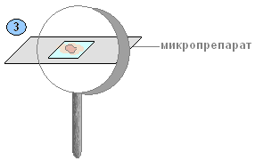
Hand magnifier
2. Consider a hand-held magnifying glass. Write the names of its parts and the functions they perform.
3. Take pieces of tomato pulp (watermelon, apple). Examine them with the naked eye. What do you get out?
- Answer: Soft thin peel and seeds.
4. Examine the pieces with a magnifying glass. What do you see?
- Answer: Pulp cells.
5. Conclusion
- Answer: The magnifying glass is so strong that you can see cells that are not visible to the naked eye.
Light microscope
1) Examine the microscope. Find the main parts of the microscope. Using the textbook text and drawing, find out what their meaning is.
2) Familiarize yourself with the rules of working with a microscope. Learn to set the light, achieve good illumination of the field of view.
3) Check each other’s knowledge of the rules for using a microscope.
4) Determine how many times the microscope magnifies the image of the object. (300 times. Depends on the microscope)
5) Practice the sequence of actions when working with a microscope.
Please write a conclusion about a piece of fruit pulp under a magnifying glass
- Even with the naked eye, or even better under a magnifying glass, you can see that the flesh of a ripe watermelon consists of very small grains, or grains. These are cells - the smallest “building blocks” that make up the bodies of all living organisms.
If you examine the pulp of a tomato or watermelon with a microscope magnifying approximately 56 times, round transparent cells are visible. In apples they are colorless, in watermelons and tomatoes they are pale pink. The cells in the “mush” lie loosely, separated from each other, and therefore it is clearly visible that each cell has its own membrane, or wall.
Conclusion: A living plant cell has:
1. Living contents of the cell. (cytoplasm, vacuoles, nucleus)
2. Various inclusions in the living contents of the cell. (deposits of reserve nutrients: protein grains, drops of oil, starch grains.)
3. Cell membrane, or wall. (It is transparent, dense, elastic, does not allow the cytoplasm to spread, and gives the cell a certain shape.) - Even with the naked eye, or even better under a magnifying glass, you can see that the flesh of a ripe watermelon consists of very small grains, or grains. These are cells - the smallest “building blocks” that make up the bodies of all living organisms.
If you examine the pulp of a tomato or watermelon with a microscope magnifying approximately 56 times, round transparent cells are visible. In apples they are colorless, in watermelons and tomatoes they are pale pink. The cells in the “mush” lie loosely, separated from each other, and therefore it is clearly visible that each cell has its own membrane, or wall.
Conclusion: A living plant cell has:
1. Living contents of the cell. (cytoplasm, vacuoles, nucleus)
2. Various inclusions in the living contents of the cell. (deposits of reserve nutrients: protein grains, drops of oil, starch grains.)
3. Cell membrane, or wall. (It is transparent, dense, elastic, does not allow the cytoplasm to spread, and gives the cell a certain shape.) - the cells are very large
- Cells are seen better when viewed under a magnifying instrument.
Even with the naked eye, or even better under a magnifying glass, you can see that the pulp of a ripe watermelon, tomato, or apple consists of very small grains or grains. These are cells - the smallest “building blocks” that make up the bodies of all living organisms.
What are we doing? Let's make a temporary microslide of a tomato fruit.
Wipe the slide and cover glass with a napkin. Use a pipette to place a drop of water on the glass slide (1).
What to do. Using a dissecting needle, take a small piece of fruit pulp and place it in a drop of water on a glass slide. Mash the pulp with a dissecting needle until you obtain a paste (2).

Cover with a cover glass and remove excess water with filter paper (3).
What to do. Examine the temporary microslide with a magnifying glass.
What we are seeing. It is clearly visible that the pulp of the tomato fruit has a granular structure (4).

These are the cells of the pulp of the tomato fruit.
What we do: Examine the microslide under a microscope. Find individual cells and examine them at low magnification (10x6), and then (5) at high magnification (10x30).

What we are seeing. The color of the tomato fruit cell has changed.
A drop of water also changed its color.
Conclusion: The main parts of a plant cell are the cell membrane, the cytoplasm with plastids, the nucleus, and vacuoles. The presence of plastids in the cell is a characteristic feature of all representatives of the plant kingdom.
3. Using the textbook, study the structure of hand-held and tripod magnifiers. Label their main parts in the pictures.
4. Examine pieces of fruit pulp under a magnifying glass. Sketch what you see. Sign the drawings.

5. After completing the laboratory work “The design of a microscope and methods of working with it” (see pp. 16-17 of the textbook), label the main parts of the microscope in the figure.

6. In the drawing, the artist mixed up the sequence of actions when preparing a microslide. Indicate with numbers the correct sequence of actions and describe the progress of preparing the microslide.
1) Place 1-2 drops of water on the glass.
2) Remove a small piece of transparent scale.
3) Place a piece of onion on the glass.
4) Cover with a cover slip and examine.
5) Stain the preparation with iodine solution.
6) Consider.
7. Using the text and pictures of the textbook (p. 2), study the structure of a plant cell, and then complete the laboratory work “Preparation and examination of a preparation of onion scale skin under a microscope.”

8. After completing the laboratory work “Plastids in the cells of the Elodea leaf” (see p. 20 of the textbook), sketch the structure of the cell of the Elodea leaf. Write captions for the drawing.

Conclusion: the cell has a complex structure: there is a nucleolus, cytoplasm, membrane, nucleus, vacuoles, pores, chloroplasts.
9. What color can plastids be? What other substances found in the cell give the plant organs different colors?
Green, yellow, orange, colorless.
10. Having studied paragraph 3 of the textbook, fill out the diagram “Cell life processes”.
Cell activity:
1) The movement of the cytoplasm - promotes the movement of nutrients in the cells.
2) Breathing – absorbs oxygen from the air.
3) Nutrition - from the intercellular spaces through the cell membrane they come in the form of nutrient solutions.
4) Reproduction - cells are capable of division, the number of cells increases.
5) Growth - cells increase in size.
11. Consider the division diagram of a plant cell. Use numbers to indicate the sequence of stages (stages) of cell division.

12. During life, changes occur in a cell.

Use numbers to indicate the sequence of changes from the youngest to the oldest cell.
3, 5, 1, 4, 2.
How does the youngest cell differ from the oldest cell?
The youngest cell has a nucleus, a nucleolus, and the oldest one does not.
13. What is the significance of chromosomes? Why is their number in a cell constant?
1) They transmit hereditary characteristics from cell to cell.
2) As a result of cell division, each chromosome copies itself. Two identical parts are formed.
14. Complete the definition.
A tissue is a group of cells that are similar in structure and perform the same functions.
15. Fill out the diagram.

16. Fill out the table.

17. Label the main parts of a plant cell in the picture.

18. What was the significance of the invention of the microscope?
The invention of the microscope was of great importance. With the help of a microscope, it became possible to see and examine the structure of the cell.
19. Prove that a cell is a living part of a plant.
A cell can: eat, breathe, grow, reproduce. And these are signs of living things.